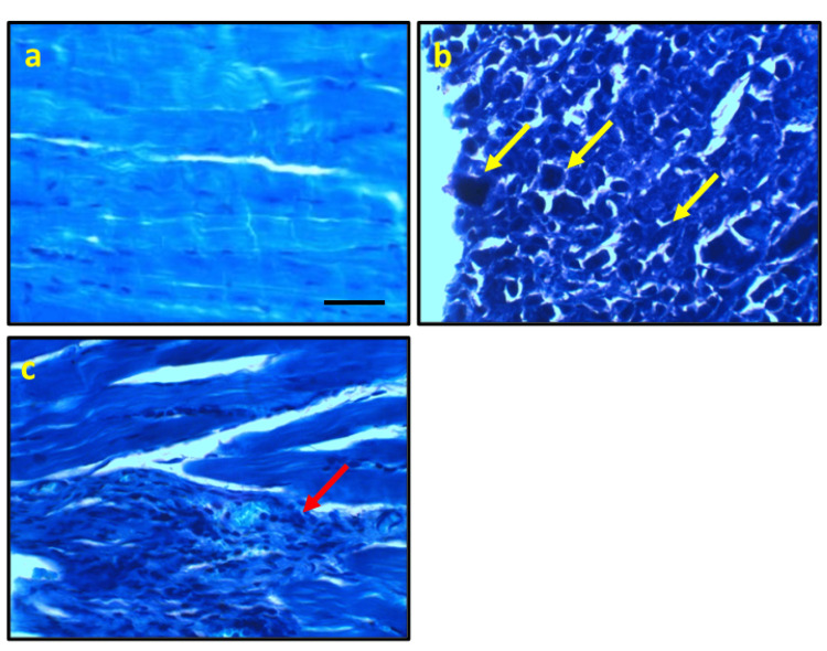
Figure 2. Representative photomicrograph stained with toluidine blue from the control group showing the normal histological appearance of muscle fiber nucleus (a), ESC showing delineated the diffuse infiltrative positive staining malignant cells with high nuclear-cytoplasmic ratio, clumped, roped, and stripping chromatin (yellow arrows, b). Ferulic acid improves the cell structure with few focal aggregations of positive stained round cells around muscle fibers (red arrows, c).
* significant difference against the control group at p<0.05. # significant difference against the ESC group at p<0.05.
Scale bar 100 μm. ESC, Ehrlich solid carcinoma.

