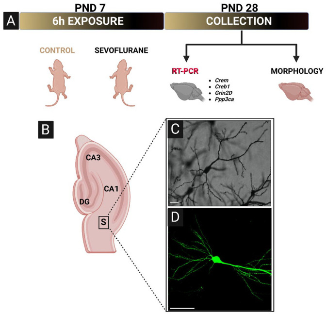
Figure 1.

Schematic representation of experimental design, imaging region of interest, and representative photomicrographs. (A) PND7 rat or Thy1-EGFP mice were exposed to 6 h of sevoflurane anesthesia. After the exposure, animals were returned to their home cages and aged to PND28 before tissue collection. A subset of rat brains was selected for gene expression analysis of four genes (Crem, Creb1, Grin2D, and Ppp3ca) critical for neuronal development, learning, and memory. The remainder of rat and all of Thy1-EGFP mouse brains were selected and processed for histomorphological analyses. (B) Schematic of hippocampal formation defining the exact region of interest used for imaging purposes. (C) Representative 20× photomicrograph of optimized Golgi-Cox impregnation of subicular pyramidal neuron in rats. Scale bar = 100 µm. (D) Representative 40× photomicrograph of subicular pyramidal neuron in Thy1-EGFP mice, showing a much better resolution of small-diameter basal and apical dendritic branching. Scale bar = 50 µm.
Crem, cAMP responsive element modulator; Creb1, cAMP responsive element binding protein 1; Grin2D, Glutamate Ionotropic Receptor NMDA Type Subunit 2D; Ppp3ca, Protein phosphatase 3 catalytic subunit alpha, a subunit of calcineurin; S, subiculum; PND, postnatal day; EGFP, enhanced green fluorescent protein.
