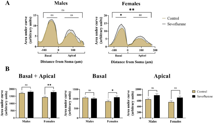
Figure 7.
Summary measurement of differences in branching complexity of apical and basal dendritic arbors with regard to sex in Thy1-EGFP expressing mouse pyramidal neurons. (A) Visual inspection of Linear Sholl Plots, plotted as a function of distance from soma, suggested upward deflection following neonatal sevoflurane treatment. However, quantitative analysis revealed no significant differences in males (Left panel). In contrast, sevoflurane-treated females (Right panel) exhibited significant upregulation of overall Sholl plot, primarily in the basal dendritic arbor. Basal dendritic arborization was arbitrarily assigned negative values on the x-axis to differentiate it from the apical arbor. (B) Quantitative analysis of AUC revealed significantly higher values only in females following neonatal sevoflurane (Left panel). When basal arbor was analyzed in isolation, neonatally exposed females had significantly increased AUC values, whereas no differences were noted in males (Middle panel). Although the trend toward higher values was observed following sevoflurane in both males and females, no statistical significance was reached in the apical dendritic arbor (Right panel). *p < 0.05; **p < 0.01.
EGFP, enhanced green fluorescent protein.

