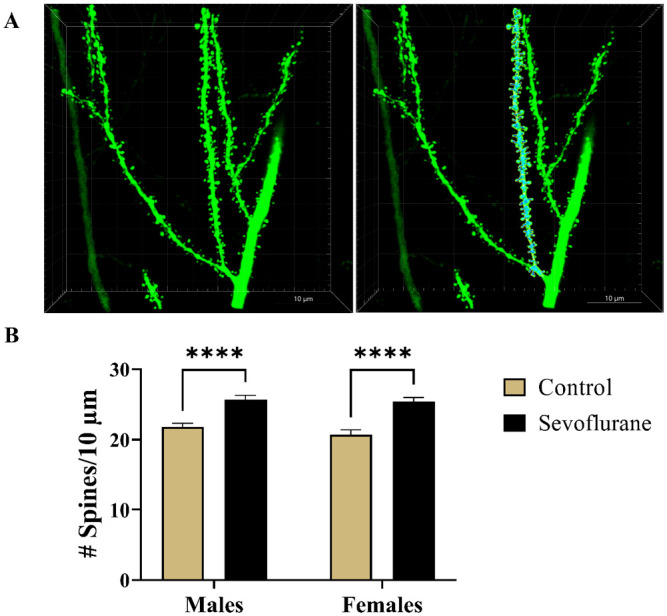
Figure 8.

Spine density analysis of Thy1-EGFP-positive apical dendritic segments of subicular pyramidal regions in mice. (A) Representative photomicrographs of second generation dendritic branches covered with spines. Corresponding raw (Left panel) and the 3D-reconstructed (Right panel) images of dendritic spines are shown at 240× magnification. (B) Analysis of dendritic spine density, expressed as the number of spines per 10-µm long branch segments, revealed highly significant 18% and 23% increase in spine density in sevoflurane-exposed males and females, respectively. ****p < 0.0001.
EGFP, enhanced green fluorescent protein.
