
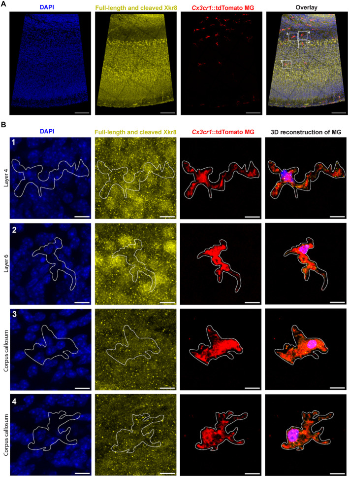
Figure EV1. Immunolabeling of Xkr8 in Cx3cr1::tdTomato microglia.
-
A, BImmunofluorescence labeling of total Xkr8 (yellow) within tdTomato+ microglia (red) in the S1 cortex of P0 mouse in mosaic image (A) and enlarged representative cells in layer 4 (B, 1), layer 6 (B, 2) and corpus callosum (B, 3 and 4); scale bars 100 μm (A) and 10 μm (B).