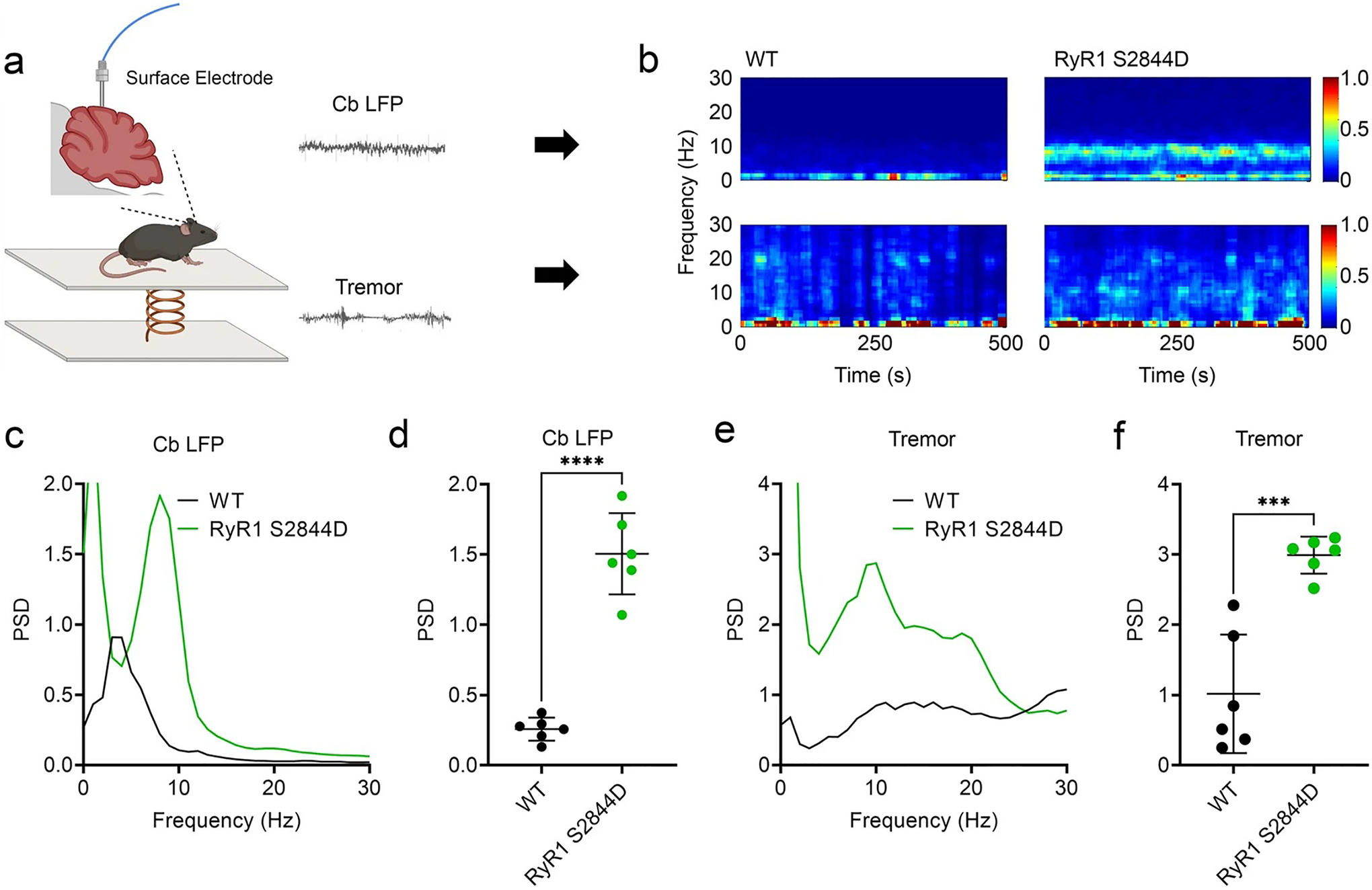
Fig. 7.

Altered cerebellar physiology in RyR1-S2844D+/+ mice. a Scheme showing simultaneous recordings of tremor and cerebellar local field potentials (LFPs) of lobules V and VI in a freely moving mouse. b Representative time–frequency plots and c, d quantified corresponding spectral diagrams of cerebellar LFP in wild-type (WT) and RyR1-S2844D+/+ mice, with a significant cerebellar LFP spike at 10 Hz in the RyR1-S2844D+/+ mice compared to WT. e A quantified spectral diagram of tremor in a WT mouse and a RyR1-S2844D+/+ mouse, f with significant tremor at 10 Hz in a RyR1-S2844D+/+ mice compared to WT. (n = 3 mice at 6–12 months old for each genotype, two independent runs for each mouse). **p = 0.0016, ****p < 0.0001 by Welch’s paired t test
