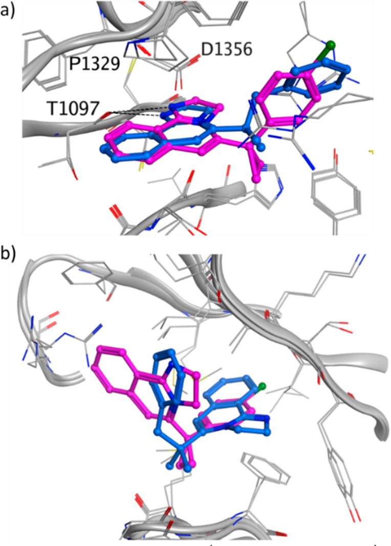
Figure 5.

Crystal structure of 3 (PDB ID: 8OG8, blue) and 4 (PDB ID: 8OG9, purple) in (a) the donut hole cavity and in (b) the blade region. For 3 only one of the blade poses is shown. The other pose has the tricyclic core flipped by 180°. The DCAF1 protein is shown in gray, and the hydrogen bonds are shown as dashed black lines.
