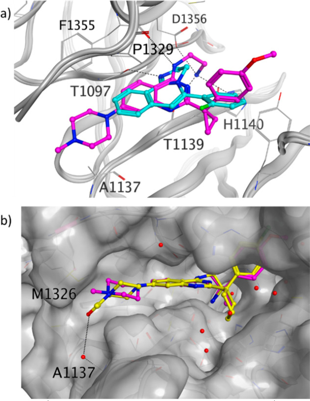
Figure 6.

(a) DCAF1 crystal structure in complex with 8 (PDB ID: 8OGB, purple) overlaid with the DCAF1 crystal structure in complex with 1 (PDB ID: 8OG6, cyan). (b) DCAF1 crystal structure in complex with 11 (PDB ID: 8OGC, yellow) overlaid with the DCAF1 crystal structure in complex with 8 (purple), of which only the ligand is shown. The DCAF1 protein surface is shown in gray, and the hydrogen bonds are shown as dashed black lines.
