Fig. 3.
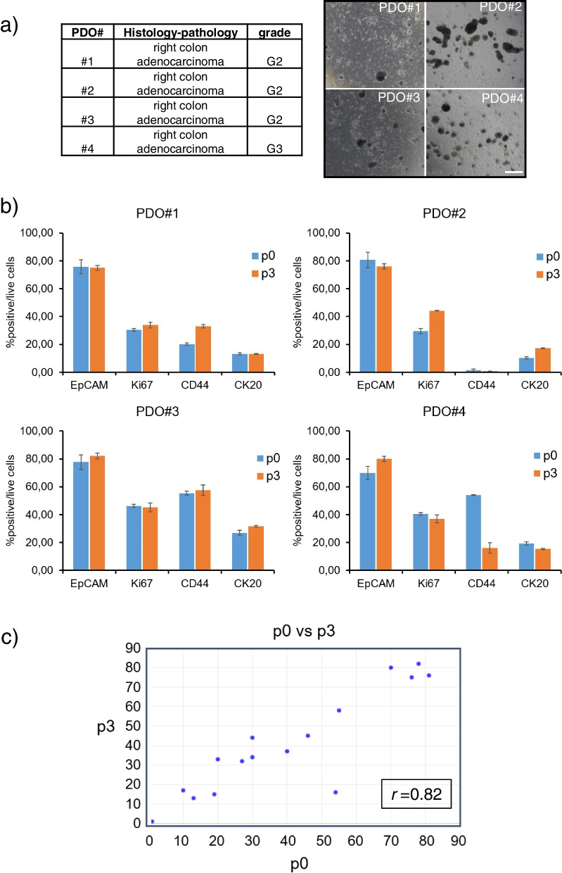
Characteristics of the CRC-derived PDOs. Patient-Derived-Organoids were obtained from four right colon adenocarcinoma specimens as described in methods. a Right panel: Clinico-pathological features of the obtained specimens. Left panel: representative micrographs of the four PDO cultures obtained from the specimens indicated in 3a, left. Size bar: 200 µm. b Validation of the PDO cultures. Upper panel: histograms showing the percentage of cells positive for EpCAM, Ki67, CD44 and CK20 in the CRC tissue immediately after the mechanical disaggregation (passage 0, p0). Lower panel: histograms showing the percentage of cells positive for the expression of the above antigens in PDO cultures disaggregated after three sequential passages (passage 3, p3). c High correlation between the number of positive cells within the p0) and the p3) specimens was shown (r = 0.8213, p < 0.01) suggesting a similar composition in cell subpopulations between the p0 and the p3 PDOs

