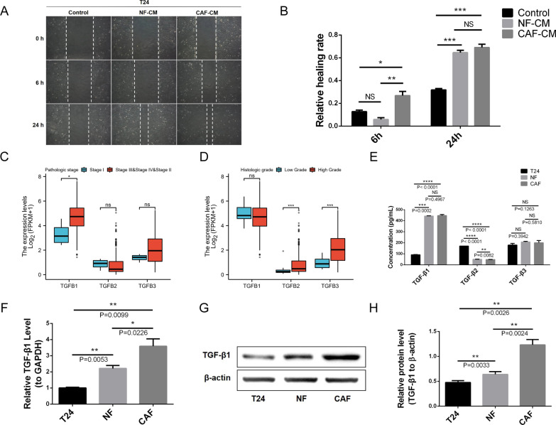
Fig. 2.
Source and expression of TGF-β1 in the tumor microenvironment. A, B CAF-CM and NF-CM promotes BLCA cell migration in a wound-healing assay. C The expression level of TGF-β1 in different pathological stages of BLCA is higher than that of TGF-β2 and TGF-β3. D The expression level of TGF-β1 in different histological grades of BLCA is higher than that of TGF-β2 and TGF-β3. E ELISA shows that the concentration of TGF-β1 in primary CAF and NF supernatant is significantly higher than TGF-β2 and TGF-β3, and that TGF-β1 derived from stromal fibroblasts is considerably higher than tumor cells. F qRT-PCR indicates that the mRNA expression levels of TGF-β1 in CAFs and NFs are significantly higher than in T24 cells. G, H WB shows that the protein expression levels of TGF-β1 in CAFs and NFs are notably higher than in T24 cells. F–H CAFs express the highest level of TGF-β1 compared to the other cell types

