
Figure 8. Extensive apoptosis in neonatal lungs.
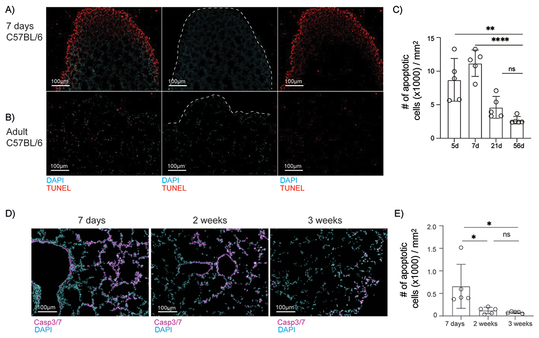
A-B. Representative images of TUNEL-stained apoptotic cells in sections from the lungs of neonatal (A) and adult (B) mice. C. Density of apoptotic cells (mean ± SD) in lung sections from mice at the indicated ages. Data are obtained from 4-5 sections per group. Significance was determined by one-way ANOVA and Tukey test for multiple comparisons. * p < 0.0332, ** p < 0.0021 D. Representative images of lung sections stained for activated caspase-3/7. Data were obtained from 5 sections per group. E. Density of apoptotic cells (mean ± SD) in lung sections from mice at the indicated ages. Significance was determined by one-way ANOVA and Tukey test for multiple comparisons.