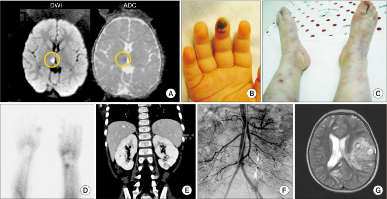
Fig. 1.
Skin manifestations and radiological findings. (A) The first magnetic resonance imaging (MRI) for brain revealed high signal intensity in diffusion weighted image (DWI), low signal intensity in apparent diffusion coefficient (ADC) on the right thalamus (yellow circle) which was conducted when the 1st neurologic symptoms; truncal ataxia, facial palsy with ptosis on the left side occurred. (B) Digital gangrene was observed on right 3rd finger. (C) Retiform purpura and livedo reticularis were shown on both lower legs with coldness. (D) Raynaud scan by 99mTc showed defects of digital blood flow on both hands, especially right the 2nd to 4th fingers and left 3rd and 4th fingers. (E) On computed tomography, several wedge-shaped focal, low attenuated lesions were observed in both kidneys and appeared to be infarctions. (F) Angiography showed multiple aneurysms in the superior mesenteric artery, inferior mesenteric artery, both renal, and splenic artery’s small branch. (G) The T2-weighted image of brain MRI showed a large amount of intracerebral hemorrhage which affects midline shifting and ventricular size at the age of 5 years.

