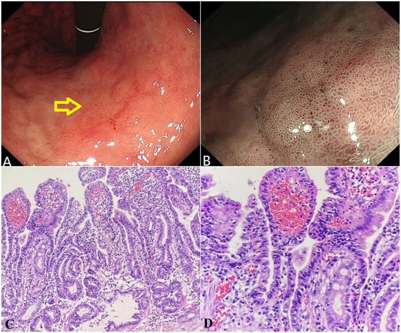
Figure 3.
Helicobacter pylori infection-related well-differentiated adenocarcinoma of an Elderly male patient. (A) White light endoscopy image. The yellow arrow indicates the lesion location. (B) NBI image. (C) Well-differentiated adenocarcinoma area (HE, ×200). (D) Well-differentiated adenocarcinoma area (HE, ×400).

