Figure 4.

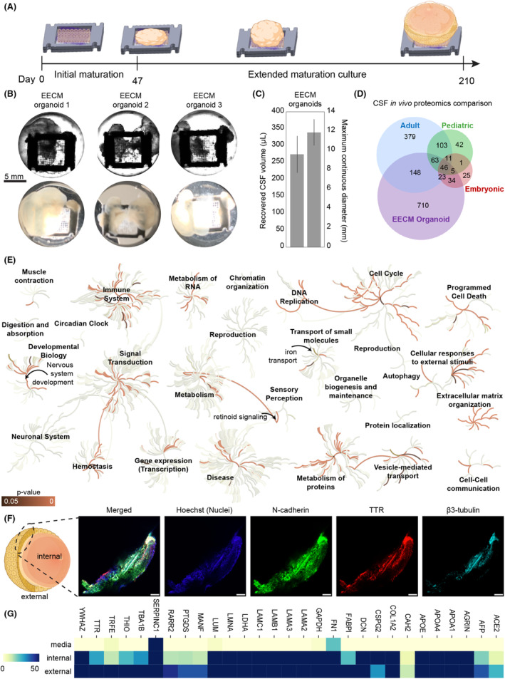
Extended organoid culture on EECMs for 7 months generated large CSF‐producing brain organoids. (A) Organoids were matured on EECMs in Midbrain Neuron Maturation Medium M2 for approximately 6 months and for a total duration of 215 days. Fluid‐filled compartments became increasingly larger over time. (B) Representative images were taken of three different EECM brain organoids using an IX83 Olympus microscope using phase contrast (top panel) and with a 12‐megapixel wide‐angle camera (bottom panel). Fluid‐filled compartments were punctured with a 22G needle and extracted for analysis. (C) Quantification of recovered cerebrospinal (CSF)‐mimicking fluid from the organoids and maximum continuous organoid diameters (n = 3 organoids). (D) Mass spectrometry was performed on the CSF fluid recovered from the organoids and compared to published proteomics signatures. 26 , 73 , 74 , 75 Values shown are for at least two organoids exceeding a relative abundance emPAI ≥ 1. (E) We used the Reactome database 38 to visualize the pathway and biological process coverage and significance from the recovered CSF‐like fluid from EECM‐supported organoids; the orange color indicates p‐value significance of protein coverage of pathway (dark = p ≤ 0.05, bright p ~ 0); gray color represents the binary status of coverage of pathway (lightest = not covered, darkest = covered). (F) Confocal images from EECM organoid section after 215 days of culture stained for N‐cadherin, TTR, and β3‐Tubulin. The scale is 200 μm. (G) Relative abundance (emPAI values) of selected proteins detected in media and internal and external organoid cultures represented as a color heatmap of lower (yellow, 0) to higher (blue, ≥ 50) relative abundance. Samples were extracted from three organoids per condition.
