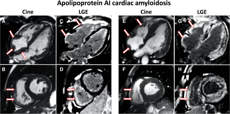
Figure 2.
Cardiac magnetic resonance images of patients with Apo AI cardiac amyloidosis. A, 4-chamber cine image acquired with steady-state free precession sequence demonstrating prominent right atrial thickening (arrows). B, Short axis cine image demonstrating prominent right ventricular thickening (arrows). C, 4-chamber late gadolinium enhancement (LGE) image acquired using phase-sensitive inversion recovery sequence reconstructions with steady-state free precession read-outs, demonstrating prominent right atrial and right ventricular LGE (arrows). D, Short axis LGE image demonstrating prominent right ventricular LGE (arrows). E, 4-chamber cine image demonstrating prominent right atrial (arrows) and right ventricular thickening (arrows). F, Short axis cine image demonstrating prominent right ventricular thickening (arrows). G, 4-chamber LGE image demonstrating prominent right atrial and right ventricular LGE (arrows). H, Short axis LGE image demonstrating prominent right ventricular LGE (arrows).

