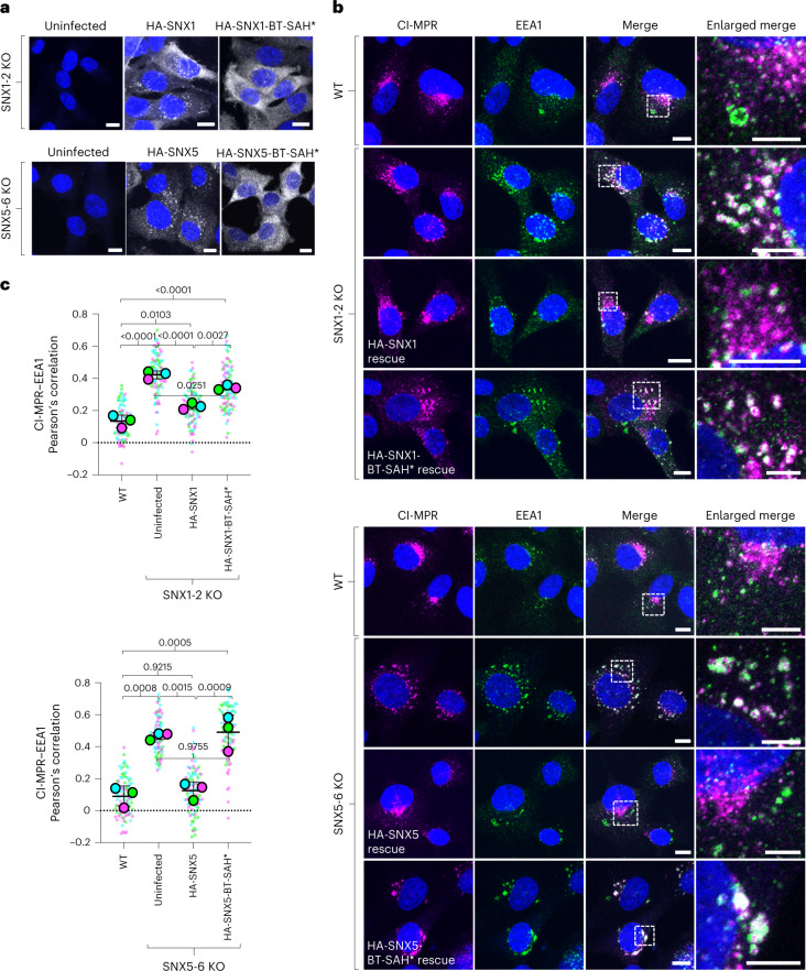
Fig. 6. Requirement of BARtip-to-BARtip and BARtip-to-PX interactions for the function of SNX1 and SNX5 in the export of CI-MPR from endosomes.
a, Immunofluorescence microscopy of fixed-permeabilized double SNX1-2 KO and double SNX5-6 KO HT1080 cells stably transduced with HA-tagged WT or BT-SAH* mutant SNX1 or SNX5. Cells were immunostained for the HA epitope and nuclei (DAPI; blue). Scale bars, 10 μm. The experiment was repeated twice with similar results. b, WT, untransduced and stably transduced double KO HT1080 cells were immunostained for the CI-MPR (magenta), early endosomes (green) and nuclei (blue), and examined by confocal fluorescence microscopy. Scale bars, 10 μm. Enlarged views of the boxed areas in the merged images are shown on the right column. Scale bars, 5 μm. c, PCC of colocalization between CI-MPR and EEA1 from experiments such as that shown in b. PCCs were calculated for 30 cells in each of three independent experiments. Data are represented as SuperPlots showing the individual data points in each experiment, the mean from each experiment and the mean ± s.d. of the means. Statistical significance was analyzed by one-way ANOVA with multiple comparisons using Tukey’s test; P values are indicated on the plots.

