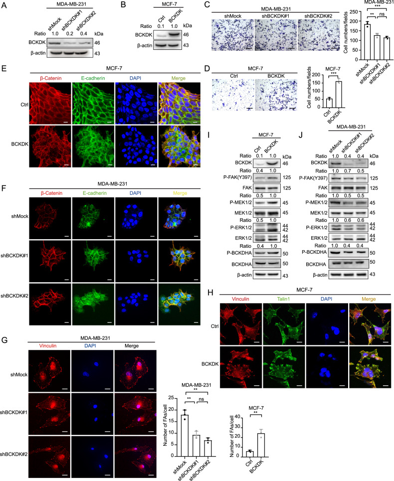
Fig. 2. BCKDK was able to promote dissociation of AJs, FA formation, and cell migration in breast cancer cell lines through inducing FAK/MAPK signal pathway.
A BCKDK was overexpressed in MCF-7 cell lines. B BCKDK was knocked down in MDA-MB-231 cell lines. C, D The migration capability of MCF-7 and MDA-MB-231 cells was detected by transwell assay; data were represented as means ± SD of triplicate experiments. *, means P < 0. 05, **, means P < 0. 01, ***, means P < 0. 001. Scale bar: 100 μm. E, F The integrity of AJs was visualized by IF assay in both cells. AJs were visualized by E-cadherin (green) and β-catenin (red). Nuclei were counterstained with DAPI (blue). Scale bar: 20 μm. G, H The number of focal adhesions in both cells was visualized by the IF assay. FAs were visualized by Vinculin (red) and talin1 (green). Nuclei were counterstained with DAPI (blue). Scale bar: 20 μm. I, J The expression of FAK/MAPK signal pathway markers was detected by western blot.

