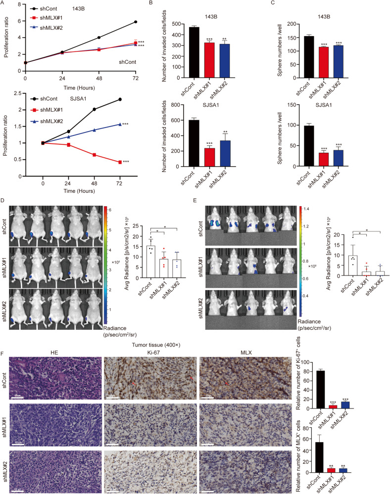
Fig. 2. MLX is essential for the growth and metastasis of osteosarcoma cells in vitro and in vivo.
A Relative growth curves of 143B and SJSA1 cells stably transduced with nontargeting scrambled control shRNA (shCont) or two MLX shRNAs (shMLX#1 and shMLX#2). Data are represented as mean ± SD, n = 3. Unpaired one-way ANOVA test followed by Dunnett’s test was used. B Invasion assay conducted in shMLX versus shCont in 143B (upper panel) and SJSA1 (lower panel). Data are represented as mean ± SD, n = 3. Unpaired t-test was used. C Sphere formation assay in 143B (upper panel) and SJSA1 cells (lower panel). Data are represented as mean ± SD, n = 3. Unpaired t-test was used. D Representative images of tumor growth in 143B-derived orthotopic model (n = 5 per group). Unpaired t-test was used. E The representative images of metastasis in 143B-derived orthotopic model (n = 5 per group). Unpaired t-test was used. F Representative images of H&E, Ki-67 and MLX staining of 143B-shCont and 143B-shMLX tumors and the respective analyses (scale bar = 50 μm). Data are represented as mean ± SD, n = 3. Unpaired t-test was used. ***: P ≤ 0.001, **: P ≤ 0.01, *: P ≤ 0.05, n.s.: not significant.

