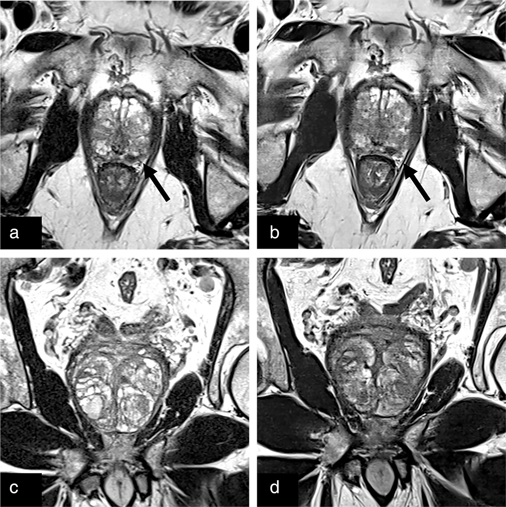
FIGURE 2:

Conventional (a) and deep learning reconstructed (b) axial T2-weighted images of the prostate with an equally well-visualized T2 hypointense lesion in the left posterolateral prostate (black arrow) later biopsied to be a grade group 2 lesion. The adjacent neurovascular bundles can be seen on both clinical and deep learning reconstructed images. Clinical (c) and deep learning reconstructed (d) coronal T2 turbo spin echo with clear prostatic capsule and BPH capsules.
