FIGURE 3.

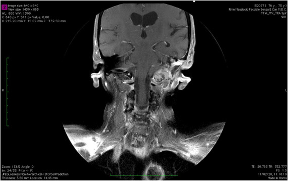
Sixteen months MRI follow up (coronal view). In the site of previous left petrosectomy the MRI scan showed an heterogeneously enhanced tissue; toward the rear, it grows through the foramen lacerum involving the foramen magnum.

Sixteen months MRI follow up (coronal view). In the site of previous left petrosectomy the MRI scan showed an heterogeneously enhanced tissue; toward the rear, it grows through the foramen lacerum involving the foramen magnum.