
FIGURE 1.
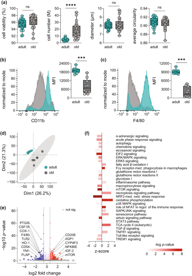
Aging establishes a distinct macrophage phenotype in the peritoneal cavity. (a) Number, viability, diameter, and circularity of isolated cells from the peritoneal cavity of adult (4–6 months) and old mice (>24 months) were assessed with a Vi‐CELL XR system (n = 25). Expression of surface markers (b) CD11b and (c) F4/80 on PM from adult (turquoise) and old mice (grey) was measured by flow cytometry (n = 9–10). Results are shown in representative histograms for both surface markers with mean fluorescence intensity (MFI) of all replicates. (d) Principal component analysis (PCA) of the proteome of PM from adult and old mice measured by DIA mass spectrometry (n = 5). (e) Volcano plot displays proteins significantly increased or decreased by aging (Table S1; n = 5). Not affected proteins are shown in grey. Dashed lines indicate a cutoff for significance of p < 0.05 and absolute fold changes (log2) > 0.58, respectively. Significantly regulated proteins of interest were labelled with an emphasis on those involved in macrophage function during inflammation. (f) Ingenuity pathway analysis of significantly regulated protein clusters in PM from old mice in comparison with adult (n = 5). Displayed pathways are among the Top 100 most significantly regulated pathways and were selected based on the relevance for aging and inflammation. Bonferroni–Holm corrected p‐values are implicated by color and z‐scores by bar size. Statistics: Data are shown as (a–c): median (min to max) or (e, f) median and p‐values were calculated by ‘a–c’ unpaired two‐tailed Student's t‐test with or without Welch's correction (Table S7), (e) Spectronaut™ (Table S1) or (f) QIAGEN Ingenuity Pathway Analysis. ***p ≤ 0.001, ****p ≤ 0.0001, ns, not significant.