
FIGURE 2.
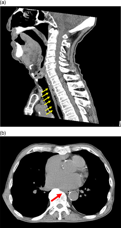
Sagittal computed tomography reveals thickening of the entire esophageal wall with an upper esophageal predominance (a). Horizontal computed tomography shows compression of the esophagus by the left atrium (b).
Official websites use .gov
A
.gov website belongs to an official
government organization in the United States.
Secure .gov websites use HTTPS
A lock (
) or https:// means you've safely
connected to the .gov website. Share sensitive
information only on official, secure websites.
Sagittal computed tomography reveals thickening of the entire esophageal wall with an upper esophageal predominance (a). Horizontal computed tomography shows compression of the esophagus by the left atrium (b).