
Figure 1. Two Hearts Depicting the Variability In Both Extension and Depth Of Trabeculae and Recesses.
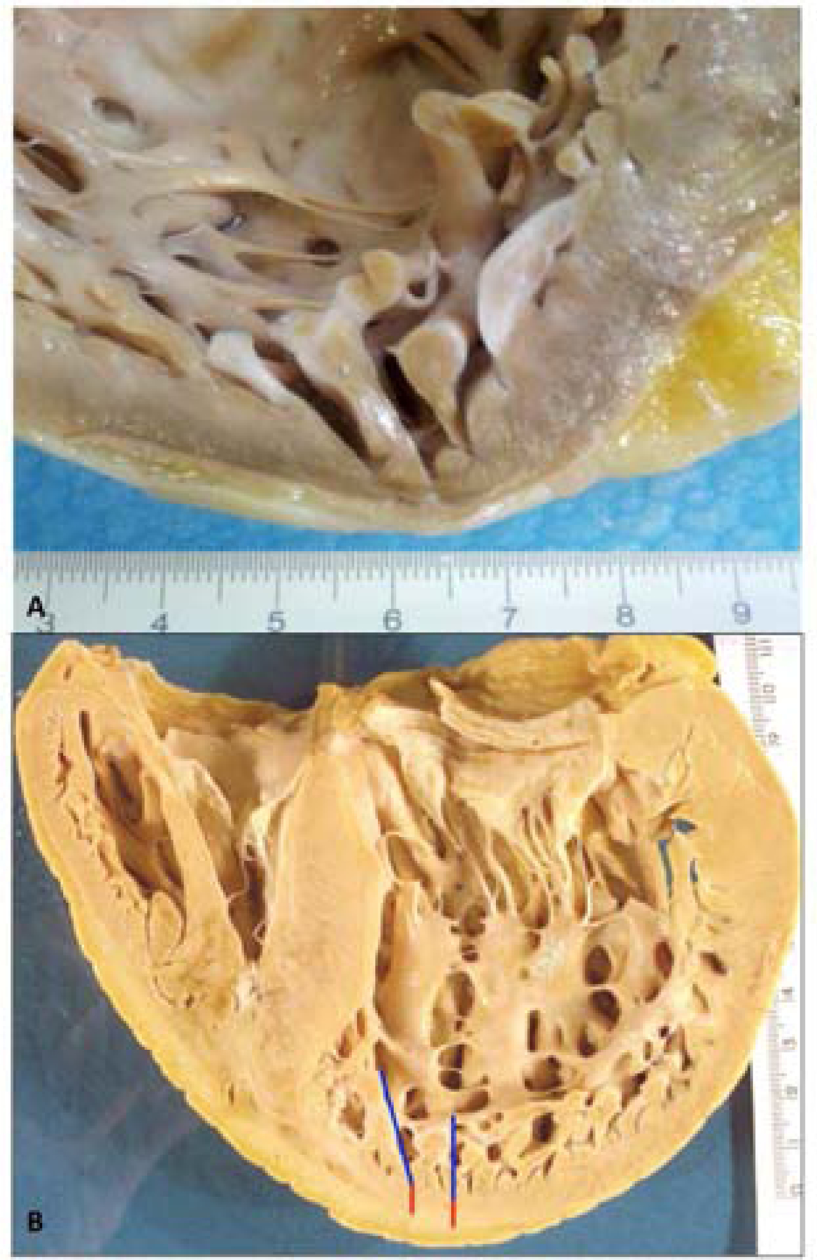
A) In this high magnification view of the apical wall of the heart, the noncompacted area is limited to a few apical trabeculae. The patient harbored mutations p.(Arg495Trp) in Myosin Binding Protein Cardiac 3 (MYBPC3) and p.(Asp117Asn) in Lim domain binding protein 3 (LDB3) genes [MH+D OH GAD EG-MYBPC3[p.Arg495Trp]+LDB3 [p.Asp117Asn]SC-IV]. Although LBD3 is a candidate gene for LVNC, in this family, the disease segregated with the mutation in MYBPC3.
B) In this heart, the prominent trabeculations (blue line) and deep recesses (red line) involve the entire LV apex. LV = left ventricle.