
Figure 3. Echocardiographic 4-chamber views distinguishing prominent trabeculation (A) vs. hypertrabeculation (B).
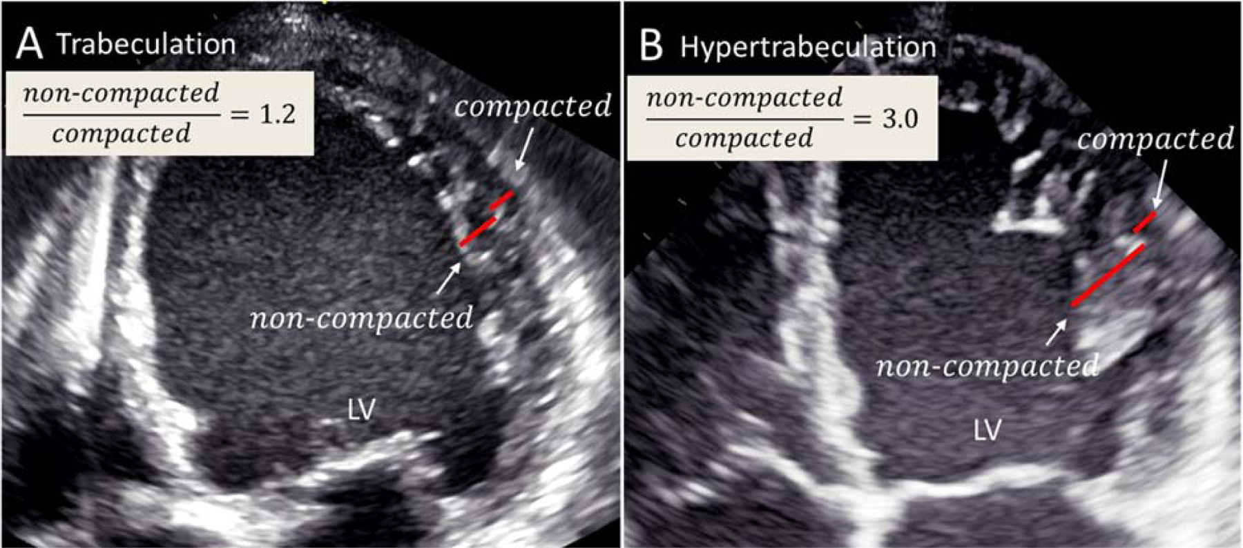
(A) An echocardiographic 4-chamber view from a patient with a dilated cardiomyopathy presenting with prominent trabeculation in the LV apex and lateral wall. In this case the criteria for LVNC are not fulfilled. (B) An echocardiographic 4-chamber view from a patient with a typical LVNC presenting with hypertrabeculation in the LV apex and lateral wall. LV = left ventricle; LVNC left ventricular noncompaction.