
Figure 4. A) An echocardiographic image from a patient with LVNC.
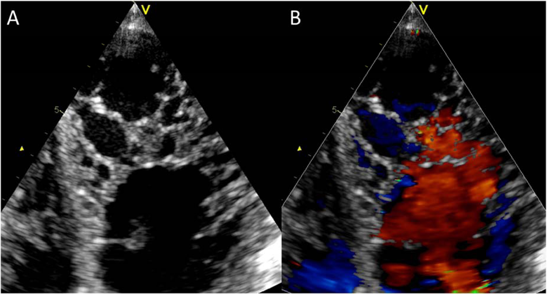
An atypical 4-chamber view was used to better illustrate the non-compaction in the LV apex. B) The same view with color Doppler imaging. This view highlights perfusion of intertrabecular recesses from the left ventricular cavity. Abbreviations as in Figure 3.