
Figure 5. Cardiac magnetic resonance (CMR) from a patient with ischemic heart disease and ejection fraction = 27%.
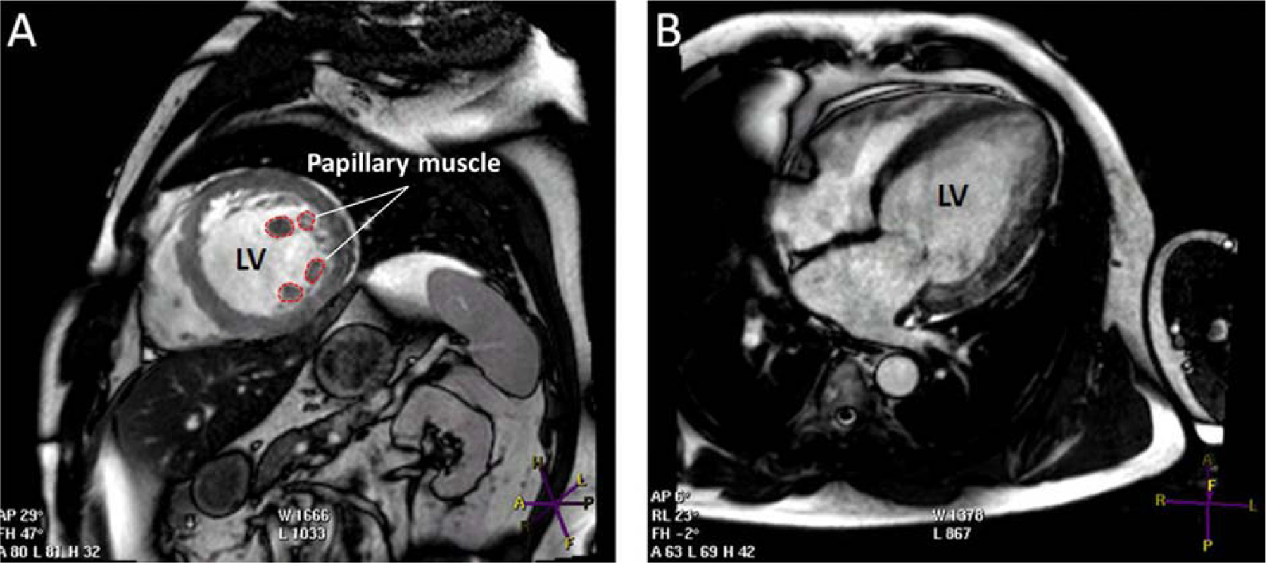
Apart from the ischemic heart disease history, this patient does not meet the CMR criteria for LVNC cardiomyopathy. A) Short axis view showing the papillary muscle with prominent trabeculation in mid left ventricle segments. B) Long axis view showing trabeculation mainly in left ventricular lateral segments. Other abbreviations as in Figure 3.