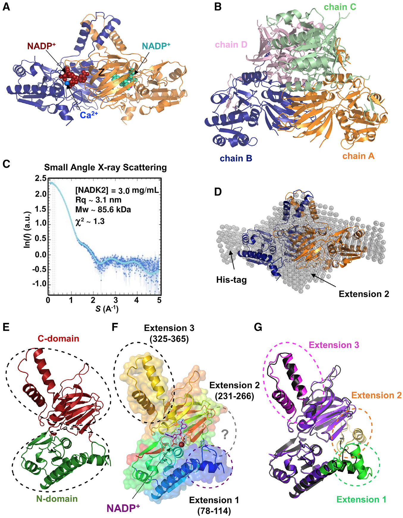
Figure 1. Crystal structure of NADK2.

(A) Crystal structure of the NADK2 in complex with NADP+ at 2.3 Å. The dimeric structure of NADK2 as deduced from crystal symmetry is shown with chain A in blue ribbon and its crystal symmetric unit, chain B, in orange. NADP+ (red or cyan in different monomers) and bound calcium (blue sphere) are shown.
(B) Crystal structure of the tetrameric cytosolic NADK. The crystal structure of cytosolic human NADK deposited in PDB (PDB: 3PFN) in the same orientation as the crystal structure of NADK2 (shown in A). The four subunits (chains A–D) of cytosolic NADK are shown as ribbons and colored by chains (A, orange; B, blue; C, light green; D, light pink).
(C) SAXS profile of purified NADK2. SAXS experimental data were fitted to a theoretical SAXS curve that was computed from the NADK2 crystal structure (dimeric form) completed with the Histag and the 2nd extension. χ2 ~ 1.3 was calculated with Crysol. Guinier plot of the SAXS curve indicated a radius of gyration (Rq) ~ 3.1 nm and a molecular mass (MW) of ~85.6 kDa.
(D) Crystal structure of the NADK2 fitted in the SAXS envelope. The dimeric crystal structure (same coloring schemes as in A) was superimposed onto the SAXS envelope. Arrows indicate the positions of the polyhistidine (His) tag and the second NADK2 extension (aa 231–266), which was not observed in the crystal structure but deduced from the AlphaFold structure.
(E) Crystal structure of the NADK2 monomer indicating the N-terminal domain (green) and the C-terminal domain (red).
(F) Monomeric NADK2 shown in ribbon was colored in rainbow colors depicting specific NADK2 extensions (1–3). Extension 2 (aa 231–266) is not visible in the electron density, but the deduced model from AlphaFold is shown in gray. NADP+ is shown in violet stick, with its electron density in violet surface, and calcium ion is shown as a blue sphere.
(G) Comparison of the crystal structure and the AlphaFold model of NADK2. The monomeric NADK2 crystal structure is shown in black ribbon. The AlphaFold2 model is colored in violet ribbon, and the three extensions are indicated in green, brown, and pink.
