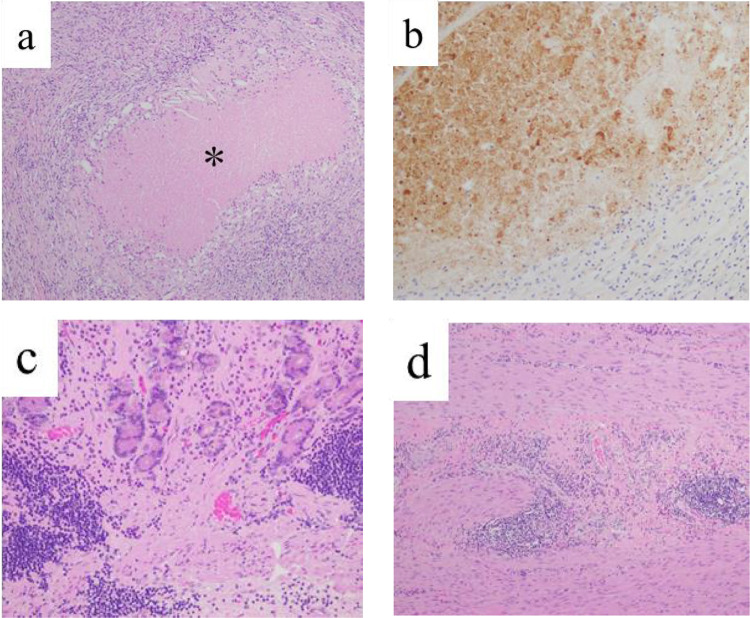
Figure 5.
Histopathological findings of Virchow’s lymph nodes and primary gastric cancer by the resected specimen. (a) On hematoxylin and eosin staining of the resected Virchow’s lymph nodes, there is coagulation necrosis (*) surrounded with histiocyte. There are no viable cancer cells. (b) On immunohistochemical staining for cytokeratin (clone AE1/AE3) of the resected Virchow’s lymph nodes, the necrotic regions were stained, indicate of necrotic change of cancer cells. (c, d) On hematoxylin and eosin staining of the resected stomach, mucosa (c) and muscularis propria (d) failed to show any viable cancer cell.

