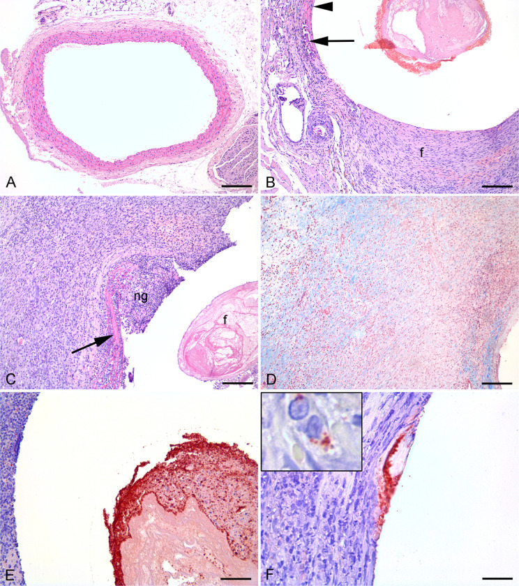
Fig 5. Histological and immunohistochemic images of infected and sterile graft, artery and sorrounding tissue from rats 19 days after insertion.
A: Normal left a. carotis (HE), bar = 250 μm. B: Right a. carotis 19 days after insertion of a vascular graft. Neointima (ni) formation, tunica media atrophy (arrow) and fibrosis of tunica adventitia (HE), bar = 100 μm. C-F: Right a. carotis 19 days after insertion of a vascular graft and inoculation with Staphylococcus aureus. C: Pink fibrin (f) is present within graft lumen. Neutrophil granulocyte (ng) infiltration, broken elastic bands (arrow) and surrounding macrophages and collagen producing fibroblasts (HE), bar = 200 μm. D: Masson trichrome staining to identify collagen in blue, bar = 200 μm. E: Red S. aureus positive bacteria located on fibrin within graft lumen (IHC), bar = 100 μm. F: Red S. aureus positive bacteria located on the vessel surface towards the graft. Insert shows red positive bacteria within a macrophage (IHC), bar = 80 μm.

