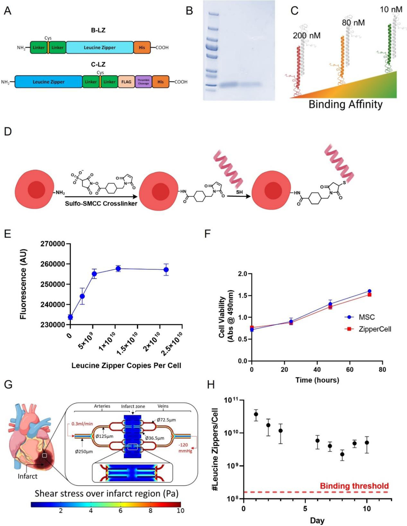
Figure 2.

Design of leucine zippers and ZipperCell conjugation. (A) Structures of customized synthetic leucine zipper constructs. (B) Coomassie Brilliant Blue stained SDS polyacrylamide gel with ladder, complementary leucine zipper (C-LZ) 10nM, and base leucine zipper (B-LZ), respectively. (C) Three orthogonal pairs of leucine zippers with varying binding affinities of 10, 80, and 200 nM. (D) Schematic representation of cellular leucine zipper decoration via heterobifunctional crosslinker (Sulfo SMCC), followed by maleimide-thiol conjugation. (E) Leucine zipper density on cells is controlled by varying the leucine zipper concentration. (F) Viability of ZipperCells. (G) Computational model in COMSOL Multiphysics to calculate the required leucine zipper densities for a stable network under cardiac flow conditions. (H) Detection of leucine zippers on cells as a function of time. Data are presented as mean ± SD with *p <0.05, **p<0.01, ****p<0.0001 by one-way ANOVA followed by Tukey’s post-test, n=3.
