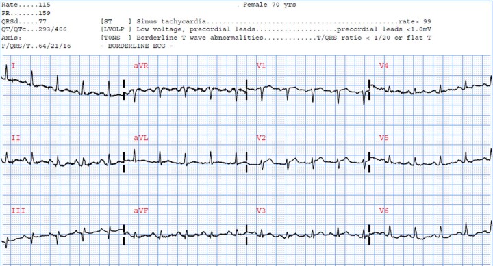
Extended Data Fig. 5. Selected example of a missed OMI by our model.
This figure provides a selected example of a patient with occlusion myocardial infarction that was missed by the machine learning model and other reference standards. This ECG was obtained on a 70-year-old female with a past medical history of hypertension, high cholesterol, prior myocardial infarction, and current smoking. The baseline clinical interpretation suggests normal sinus rhythm with benign findings. There are isolated Q waves in inferior leads, low ECG voltage, and some baseline wander and high frequency noise in few leads. The OMI risk score was 2 indicating a low risk. The patient was later sent to the catheterization lab, which showed severe left main occlusion and had many stents placed. The patient developed new-onset HF during hospitalization. A closer look at this ECG by experienced ECG readers suggests that this ECG could resemble the ‘precordial swirl pattern’, a rightward ST-elevation vector, with STE in V1 and aVR and reciprocal ST-depression in V5 and V6. This pattern was found to correlate with LAD occlusion.

