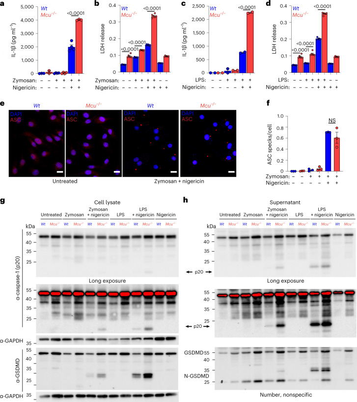
Fig. 6. Mcu−/− macrophages display increased inflammasome activation and output.
a, IL-1β enzyme-linked immunosorbent assay (ELISA) of cell supernatants collected from WT and Mcu−/− macrophages. Cells were stimulated with zymosan for 3 h followed by nigericin (5 µM) overnight. N = 3 biological replicates. Error bars represent the s.e.m.; P values were determined by one-way ANOVA. b, LDH levels in cell supernatants were collected from WT and Mcu−/− macrophages. N = 6 biological replicates. Error bars represent the s.e.m.; P < 0.0001 determined by one-way ANOVA. c, IL-1β ELISA of cell supernatants collected from WT and Mcu−/− macrophages. Cells were stimulated with LPS for 3 h followed by nigericin (5 µM) overnight. N = 2 biological replicates. Error bars represent the s.e.m.; P values were determined by one-way ANOVA. d, LDH levels in cell supernatants collected from WT and Mcu−/− macrophages. N = 6 biological replicates. Error bars represent the s.e.m.; P < 0.0001 determined by one-way ANOVA. e, Representative images of Mcu−/− and WT macrophages immunostained for ASC and nuclei (DAPI). Cells were stimulated with zymosan for 3 h followed by nigericin (5 µM) for 1 h. f, Quantification of ASC specks, average number of specks counted per cell, for WT and Mcu−/− macrophages. N = 3 biological replicates. Error bars represent the s.e.m.; no significance was determined by the one-way ANOVA. g, Western blot analysis of cell lysates from WT and Mcu−/− macrophages stimulated with zymosan for 3 h followed by nigericin (5 µM) for 1 h. Cell lysates were immunoblotted for caspase-1 (p20), GSDMD and GAPDH. h, Western blot analysis of supernatants corresponding to samples shown in g. N = 1 representative replicate, from two independent experiments. Cell supernatants were immunoblotted for caspase-1 (p20), GSDMD and GAPDH.

