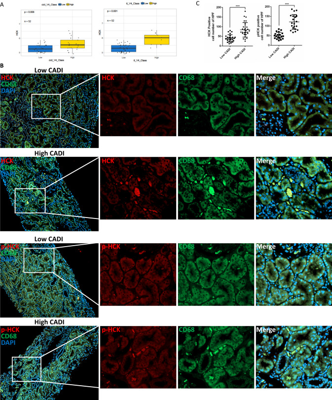
Fig. 1. HCK’s expression was associated with inflammation and fibrosis in allograft biopsies.
A The expression of HCK was up-regulated in high i + t and ci+ct scores (>=2 vs. <2) vs low scores in allograft biopsies at 12 months after transplantation. B IHC Staining of phosphorylated-HCK, HCK and CD68 in high chronic allograft damage index (CADI) and low CADI allograft kidneys. C Quantification (n = 4 biopsies/group; 5-6 random fields/biopsy) of the phosphorylated-HCK and HCK positive cells in the high and low CADI biopsies. i, interstitial inflammation; t, tubulitis. ci, interstitial fibrosis; ct, tubular atrophy. ***p < 0.001. Source data are provided as a Source Data file.

