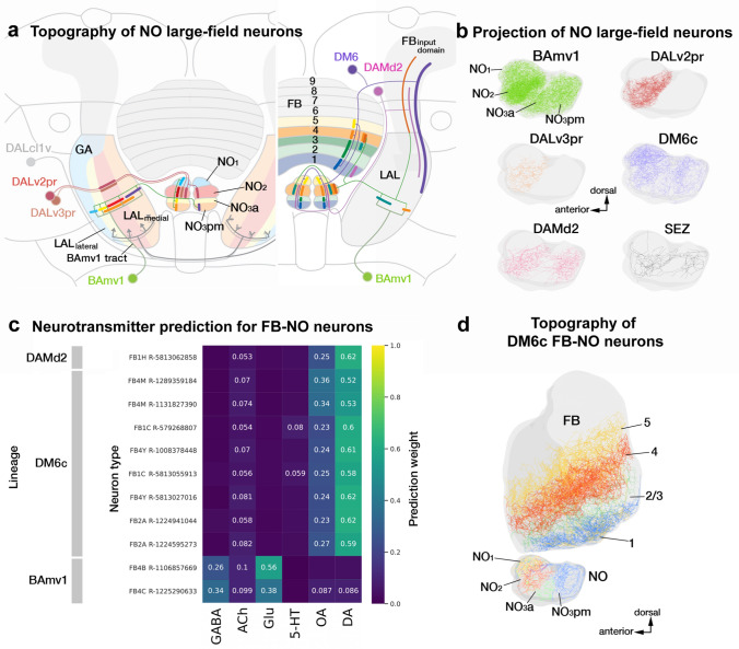
Fig. 6.
Organization of the noduli (NO) and the lateral accessory lobe (LAL). a Schematic frontal section of the brain hemisphere at the central level, visualizing the fan-shaped body (FB) and NO with surrounding neuropil compartments, as well as lineages with neurons that provide innervation of the NO. One major lineage, BAmv1 (light green), connects different vertical bands of the LAL with specific NO compartments. The position of the BAmv1 lineage-associated tract divides the LAL into a narrow lateral domain (cyan, yellow) and a medial domain (red, orange). As shown for FB large-field neurons, one detects a loose correlation between lateral-medial position of dendritic branches in the LAL and dorsal–ventral position of endings in the NO. Two neurons included in lineages DALv2pr and DALv3pr also form connections between LAL and NO. Right side of the panel (a) depicts the second major lineage, DM6, for NO input neurons. These neurons, in addition to the NO, have branches in the ventral half of the FB, as well as the FB input domain. Dorso-ventral position of the FB layer and dorso-ventral position of the connected NO compartment are strictly correlated, as indicated by corresponding colors. A small number of neurons of BAmv1 and DAMd2 also interconnect FB and NO in the manner indicated. b Plots of axonal arbors of NO-innervating neurons schematically shown in (a), presented in lateral view (anterior to the left, dorsal top). c Heatmap showing neurotransmitter predictions for different FB-NO neurons. d Plots of complete arbors of four representative DM6 FB-NO neurons in lateral view (anterior to the left, dorsal up), rendered in different colors. For abbreviations see Table 1

