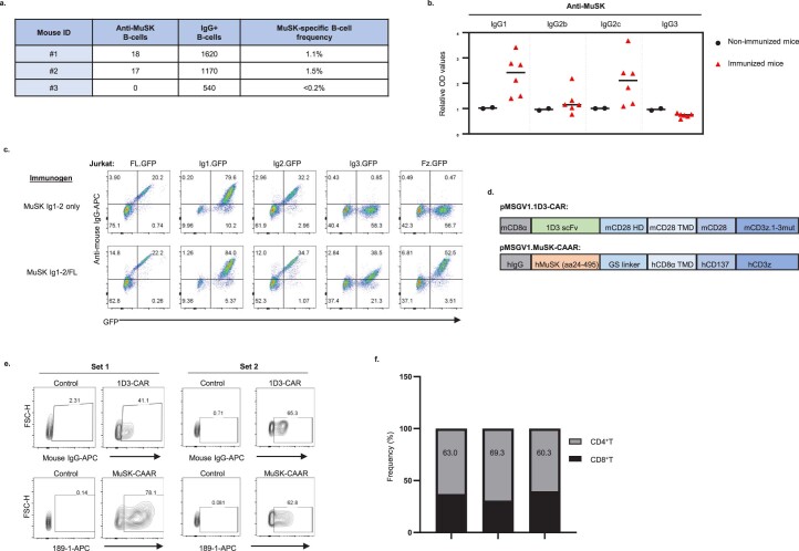
Extended Data Fig. 7. Immunologic characterization of the MuSK EAMG syngeneic MuSK-CAART treatment model.
a) Splenocytes were harvested from select MuSK-immunized mice, and purified B-cells were evaluated using a MuSK-specific and total IgG B-cell ELISpot assay to quantitate the MuSK-specific B-cell frequency. Dots from duplicated wells were summarized after normalization with seeded cells (dots per 100,000 cells). MuSK-specific B-cell frequency is calculated by dividing anti-MuSK B-cells by total IgG+ B-cells. b) IgG subclasses of anti-MuSK antibodies were detected in sera from MuSK-immunized mice in reference to sera from negative controls (non-immunized C57BL/6 and Rag2IL2rγ-deficient mice) and median value was plotted. c) Epitope mapping of serum from a mouse immunized and boosted with MuSK Ig1-Ig2, or MuSK Ig1-Ig2 followed by MuSK full length (FL) boost, using Jurkat CAAR T-cells expressing individual MuSK domain CAARs linked to GFP. Representative FACS plots are shown from three independent experiments. d) Schematic of pMSGV1-1D3-CAR (mouse CD8α signal peptide, 1D3 anti-mouse CD19 single chain variable fragment (scFv), mouse CD28 hinge (HD)/transmembrane domain (TMD)/costimulatory domain, and a mouse CD3ζ.1-3mut domain to confer enhanced persistence. Schematic of pMSGV1-MuSK-CAAR (human MuSK extracellular domains (amino acids 24-495), glycine-serine (GS) linker, hCD8α TMD, and human CD137-CD3ζ). e, f) Transduction efficiency of 1D3-CAR and MuSK-CAAR in primary mouse T-cells (Set 1 and Set 2) and g) percentage of CD4+/CD8+ T-cells in Set 1 was evaluated on day 4 after T-cell activation (one day prior to injection).

