
FIGURE 5.
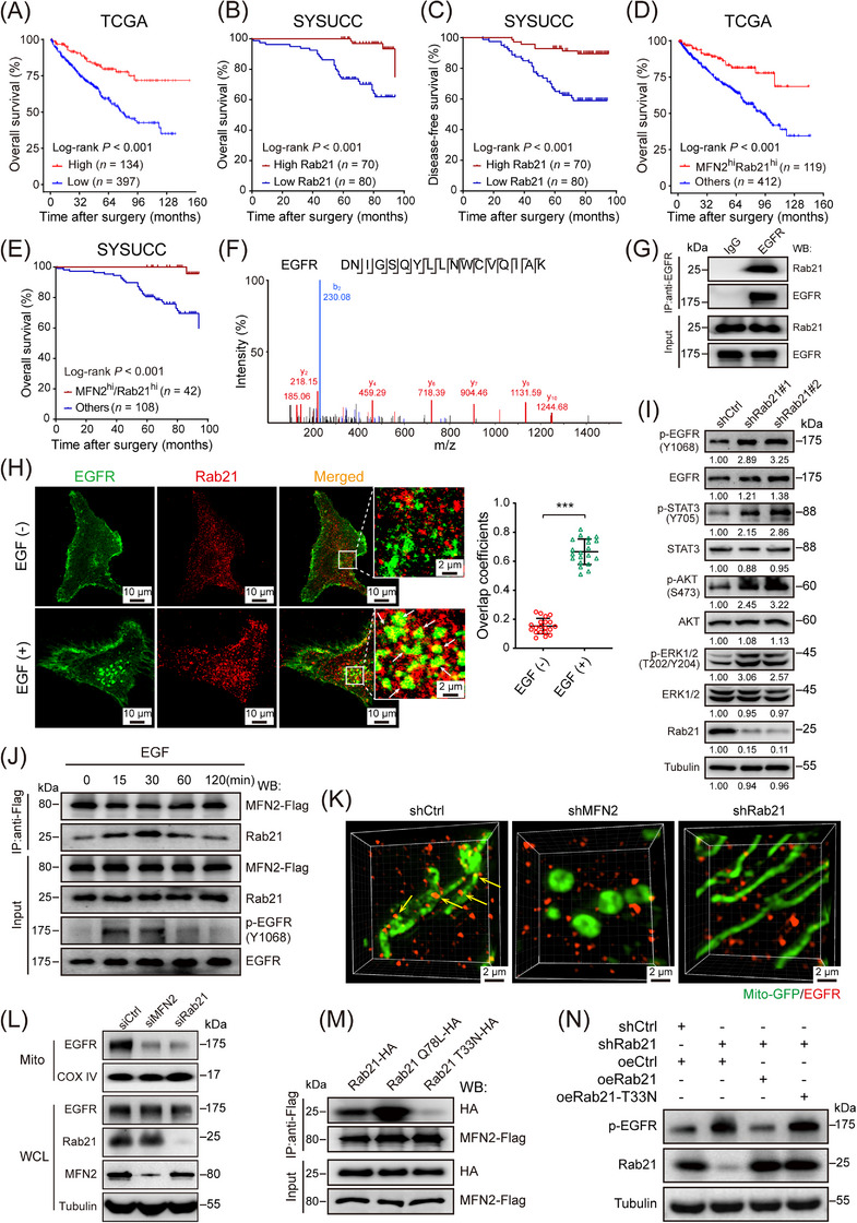
Rab21 interacts with EGFR and delivers EGFR to mitochondria in ccRCC cells. (A) OS of ccRCC patients with high (n = 134) or low (n = 397) expression of Rab21 in TCGA‐KIRC cohort. (B‐C) OS (B) and DFS (C) of ccRCC patients with high (n = 70) or low (n = 80) expression of Rab21 in SYSUCC cohort. (D) OS of ccRCC patients, with regard to the simultaneous high expression of MFN2 and Rab21 (n = 119), in TCGA‐KIRC cohort. (E) OS of ccRCC patients, with regard to the simultaneous high expression of MFN2 and Rab21 (n = 42), in SYSUCC cohort. (F) LC‐MS identified an EGFR‐unique 17‐aa peptide sequences in 786‐O cells. (G) Co‐IP of endogenous EGFR from 786‐O cells indicated that EGFR couples with Rab21. IgG was used as a control. (H) Representative immunofluorescence staining showing the co‐localization of EGFR and Rab21 in 786‐O cells upon EGF stimulation (left panel). EGF (‐): cells not treated with EGF; EGF (+): cells treated with 100 ng/mL EGF for 20 min. Statistical quantification of the co‐localization between MFN2 and Rab21 is shown in right panel. (I) Western blotting analysis of EGFR pathway proteins in Rab21‐knockdown 786‐O cells using the indicated antibodies. (J) MFN2 dynamically interacts with Rab21 in 786‐O cells. 786‐O cells were transfected with MFN2‐Flag, treated with EGF (100 ng/mL) for the indicated time points and total cell lysates were prepared. Co‐IP and the Western blotting were performed with the indicated antibodies. The interaction between MFN2 and Rab21 changed with prolonged EGF stimulation; the strongest interaction was at 30 min before declining over time. (K) Representative 3D‐SIM images of mitochondrial (mito‐GFP) localization of EGFR in the indicated 786‐O cells. (L) Mitochondrial localization of EGFR is MFN2 and Rab21‐dependent. Whole cell lysates and mitochondria were prepared from 786‐O cells with MFN2 or Rab21 silencing and then subjected to Western blotting analysis. (M) Co‐IP of MFN2‐Flag with WT Rab21, Rab21(Q78L) mutant, or Rab21(T33N) mutant indicated that MFN2 strongly interacts with the constant GTP‐loading variant Rab21(Q78L). (N) Western blotting assay of p‐EGFR levels in Rab21‐knockdown 786‐O cells with wild‐type or Rab21 mutant (T33N) reconstitution. Data are presented as means ± SD. *** P < 0.001, by Student's t test (H) or log‐rank test (A, B, C, D, E).
Abbreviations: OS, overall survival; DFS, disease‐free survival; hi, high; ccRCC, clear cell renal cell carcinoma; TCGA‐KIRC, the Cancer Genome Atlas kidney renal clear cell carcinoma; shCtrl, negative control shRNA; siCtrl, negative control siRNA; oeCtrl, empty overexpression control. LC‐MS, liquid chromatography‐mass spectrometry; WCL, whole cell lysates; Mito, mitochondria; WT, wild‐type; EGF, epidermal growth factor; IP, immunoprecipitation.