FIGURE 6.

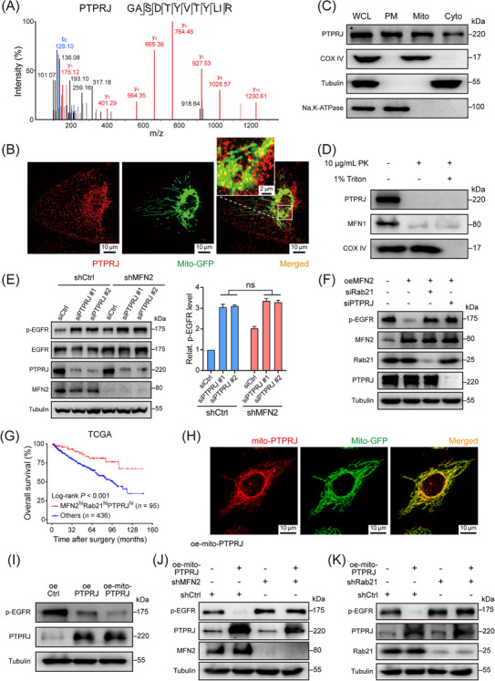
Mitochondria‐located PTPRJ dephosphorylates EGFR and inactivates EGFR signaling transduction. (A) LC‐MS analysis of mitochondria isolated from 786‐O cells identified PTPRJ as a mitochondria‐located tyrosine‐protein phosphatase. (B) Representative immunofluorescence staining image showing the co‐localization of mitochondria (green) and PTPRJ (red) in 786‐O cells stably transfected with mito‐GFP. (C) Mitochondria, cytosol and plasma membrane fractions of 786‐O cells were isolated and PTPRJ were detected by Western blotting. COX IV, tubulin, and Na, K‐ATPase were used as markers for mitochondria, cytoplasm, and plasma membrane, respectively. (D) Western blotting showing the distribution of PTPRJ on mitochondria. MFN1 and COX IV were used as markers of mitochondrial outer membrane and mitochondrial inner membrane. (E) Representative western blot image (left panel) and statistical analysis (right panel) of p‐EGFR and total EGFR protein levels using the indicated antibodies in MFN2 knockdown and control 786‐O cells transfected with PTPRJ siRNAs or control siRNA. (F) Western blotting showing p‐EGFR levels in MFN2‐overexpressing or control Caki‐1 cells with Rab21 or PTPRJ silencing. (G) OS of ccRCC patients, with regard to the simultaneous high expression of MFN2, Rab21 and PTPRJ (n = 95) in TCGA‐KIRC cohort. (H) Representative immunofluorescence staining image showing the co‐localization of mitochondria (green) and mito‐PTPRJ (red) in 786‐O cells stably transfected with mito‐GFP. (I) Western blotting showing p‐EGFR levels in 786‐O cells with PTPRJ or mito‐PTPRJ overexpression. (J) Western blotting showing p‐EGFR levels in MFN2‐knockdown or control 786‐O cells with mito‐PTPRJ overexpression. (K) Western blotting showing p‐EGFR levels in Rab21‐knockdown or control 786‐O cells with mito‐PTPRJ overexpression. Data are presented as mean ± SD. ns, not significant, by Student's t test (E) or log‐rank test (G).
Abbreviations: LC‐MS, liquid chromatography‐mass spectrometry; PTPRJ, tyrosine‐protein phosphatase receptor type J; hi, high; Mito, mitochondria; GFP, Green fluorescent protein; COX IV, cytochrome c oxidase subunit IV; Cyto, cytoplasm; PM, plasma membrane; WCL, whole cell lysate; PK, proteinase K; oe‐mito‐PTPRJ, overexpression of OMM‐anchoring form of PTPRJ; siCtrl, negative control siRNA; shCtrl, negative control shRNA; oeCtrl, empty overexpression control. OS, overall survival; Relat., relative; TCGA, the Cancer Genome Atlas.
