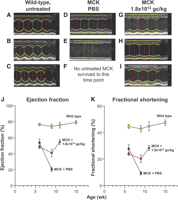
Figure 3.
Echocardiographic assessment of cardiac function of FXN-MCK mice and controls following treatment with AAVrh.10hFXN. Mice from each treatment group were monitored over time with echocardiography (see the Methods section). M-mode measurements were recorded for the short axis of the left ventricle at the midventricular level. (A–I) Representative echocardiogram tracings of the left ventricle for the same male mouse at 3 time points (7, 9, and 11 weeks of age, with the 7-week time point just before treatment and the 9- and 11-week assessments post-treatment) from each cohort. Description of the echocardiogram tracings in each panel, top to bottom: Top yellow line in each image traces the epicardial border along the anterior wall of the left ventricle, the second yellow line is a trace of the anterior endocardial wall border from full systole to full diastole. The third yellow line from the top traces the endocardial border of the posterior wall; the bottom yellow line traces the epicardial border of the posterior wall. The top two and bottom two lines together measure the wall thickness of the anterior and posterior wall of the left ventricle, respectively. The vertical red and green lines (left to right in each panel) measure the chamber of the left ventricle at full diastole and full systole, respectively. (A–C) WT untreated mouse control at 41, 64, and 76 days. (D–F) MCK mouse, PBS treated control at 42 and 63 days. (G–I) MCK mouse, 1.8 × 10¹² gc/kg dose at 42, 63, and 76 days. No mice in the PBS-treated MCK mouse cohort survived to the 11-week assessment time point. (J, K) Quantitative assessment of ejection fraction and fractional shortening calculated from the M-mode measurements of the short axis. (J) Change in ejection fraction (%) over time of WT C57Bl/6, PBS-treated, and AAVrh.10hFXN (1.8 × 10¹² gc/kg)-treated MCK mice. (K) Change in fractional shortening (%) in the same mice. Shown is the mean and standard error of percent change. All measurements were performed at 6 weeks (1 week before treatment), 9, 11, and 15 weeks of age for all surviving mice. Data from males and females were combined for each dosage cohort, shown as mean ± SEM. C57Bl/6 WT mice, yellow circles; MCK mice treated with AAVrh.10hFXN, 1.8 × 10¹² gc/kg dose, red circles; and MCK mice with PBS (controls), black circles. AAVrh.10hFXN, adeno-associated virus encoding the normal human frataxin gene; M-mode, motion mode; WT, wild type.

