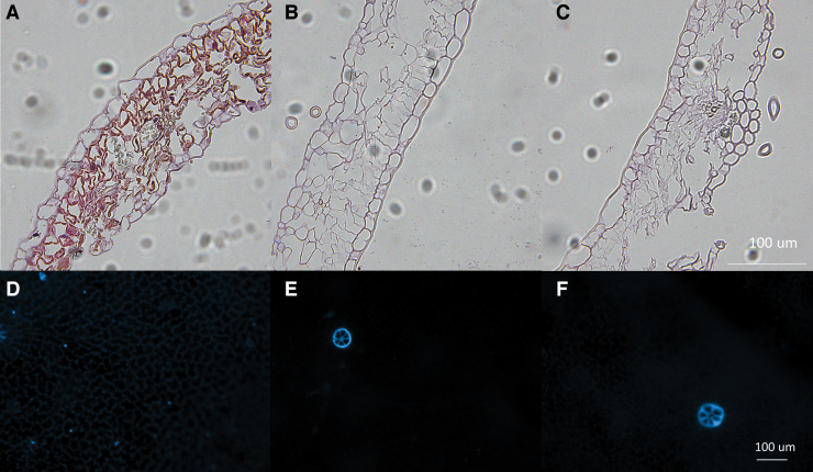
FIG. 4.
Representative H&E staining for visualization of structure of intact (A) and SDS- (B) and SDS/EGTA- decellularized (C) leatherleaf cross-section at 40 × magnification. Representative images of DAPI nuclear counterstain on the adaxial surface of leatherleaf before (D) and after decellularization with SDS (E) and SDS/EGTA (F). DAPI, diamidino-2-phenylindole; H&E, hematoxylin and eosin. Color images are available online.

