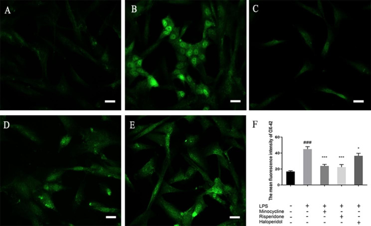
Fig. 4.
Effects of minocycline and antipsychotics on OX42 marker of microglia activated by LPS. The cells were randomly divided into 5 groups. The blank control group (A) was treated with nothing, the LPS group (B) was treated with 1.0 ug/ml LPS for 24 h. The three drug groups were pretreated with 10 μmol/L minocycline (C), 10 μmol/L risperidone (D) and 50 μmol/L haloperidol (E) respectively for 24 h, and then 1.0 ug/ml LPS was added for 24 h. The changes of microglia specific marker OX-42 were observed by immunofluorescence. Each group chose one of the three separate images as a representative. Scale bar in Fig.A is 15 μm, in Fig.B-E is 25 μm. The bar chart (F) shows statistical results of MFI. ###p < 0.001 was compared with the blank control group; *p < 0.05, **p < 0.01, and ***p < 0.001 were compared with the LPS group.

