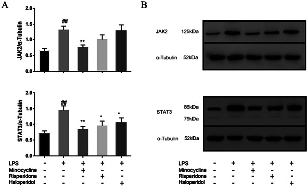
Fig. 8.
Effects of minocycline and antipsychotics on JAK2-STAT3 inflammatory signaling pathway in BV-2 microglial cells. The cells were randomly divided into 5 groups. The blank control group was treated with nothing, the LPS group was treated with 1.0 ug/ml LPS for 24 h. The three drug groups were pretreated with 10 μmol/L minocycline, 10 μmol/L risperidone and 50 μmol/L haloperidol respectively for 24 h, and then 1.0 ug/ml LPS was added for 24 h. The band density was used to quantify the expression levels of JAK2 and STAT3 normalized with a band density of α-Tubulin (A). The expression of the proteins was determined by western blot analysis (B). ##p < 0.01 was compared with the blank control group; *p < 0.05 and **p < 0.01were compared with the LPS group. The original image of the full gels/blots was included in the Supplementary Fig. S6, and Fig.S7.

