Figure S5.
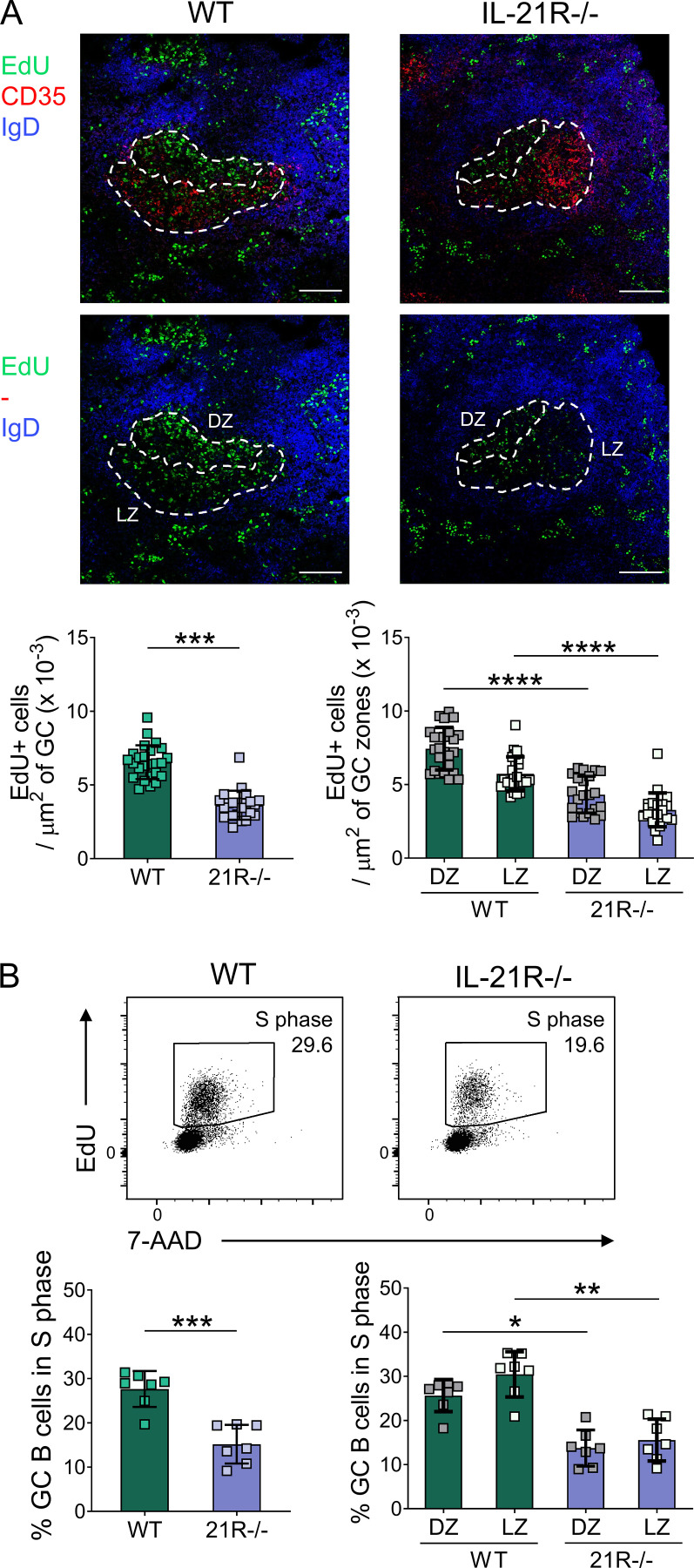
IL-21 promotes proliferation of GC B cells. Proliferating cells were analyzed in splenic GC from SRBC-immunized wild-type (WT) and IL-21R−/− mice 6 d after immunization. Mice received i.p. injections of EdU 30 min before tissue harvest. (A) Representative confocal images (top) of splenic GC and collated data (bottom) comparing EdU+ cell density in total GC and in GC dark zone (DZ; CD35−) and GC light zone (LZ; CD35+) areas from immunized WT and IL-21R−/− mice (scale bar 100 µm; 7–9 GC/mouse, each point represents a GC). Spleen sections were stained for EdU (green), CD35 (red), and IgD (blue). Data are collated from two independent experiments; n = 3; Mann–Whitney U test or Kruskal–Wallis test. (B) Representative flow cytometry plots (top) showing splenic GC B cells (CD19+Fas+GL-7+) in S phase (EdU+) and collated data (bottom) for total GC B cells and dark zone (CXCR4highCD86low) and light zone (CXCR4lowCD86high) GC B cells in S phase. Data are collated from two independent experiments; n = 7; Mann–Whitney U test or Kruskal–Wallis test. Mean ± SD are shown; ****, P < 0.0001; ***, P < 0.001**, P < 0.01; *, P < 0.05.

