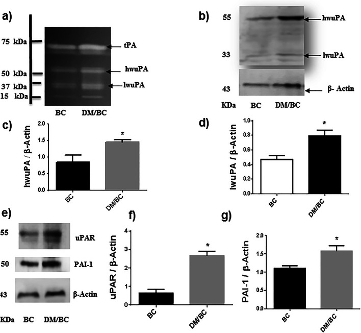
Fig. 5.
Diabetes increased the expression of components of the plasminogen activation system. The uPA was assessed by zymography in polyacrylamide gels copolymerized with casein-plasminogen (a). Additionally, uPA (b, c, and d), uPAR (e and f), and PAI-1 (e and g) were analyzed by western blot and densitometry analysis; the intensity of the bands was calculated after background subtraction and normalization to β-actin. Representative images of blots and means ± SD of three independent samples, each one obtained from 2 or 3 tumors, are presented, *p < 0.05 versus BC

