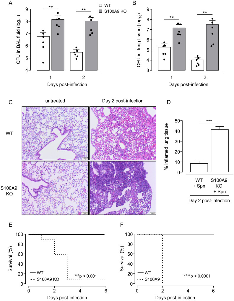
Fig 2. Effect of S100A9 deficiency on outcome in pneumococcal pneumonia.
WT mice (white bars) and S100A9 KO mice (grey bars) were infected orotracheally with S. pneumoniae and were analyzed at indicated time points. (A,B) Bacterial loads in BALF (A) and lung tissue (B) at 24 hours and 48 hours post-infection. Values are shown as mean ± SD (n = 6 mice per time point and treatment group) and are representative of two independent experiments. (C) Lung histopathology of untreated (left panels, scale bar: 100 μm) and S. pneumoniae-infected (right panels, scale bar: 100 μm) WT and S100A9 KO mice on day 2 post-infection. Illustrations (C) are representative of n = 4 mice per group. (D) Percent inflamed lung tissue of S. pneumoniae-challenged WT and S100A9 KO mice at day 2 post-infection (n = 3 mice per treatment group). (E,F) Survival analysis of WT and S100A9 KO mice challenged with a low (6 x 10⁶ CFU) (E) or increased (107 CFU) infection dose of S. pneumoniae (n = 9 mice per group). **p ≤ 0.01; ***p ≤ 0.001, ****p ≤ 0.0001 compared to WT mice (Mann-Whitney U test, t-Test, log-rank test).

