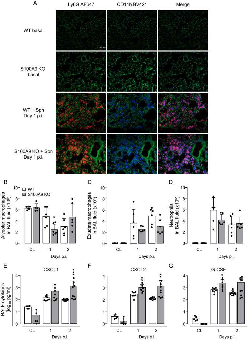
Fig 3. Leukocyte recruitment and cytokine expression in lungs of S. pneumoniae-challenged WT and S100A9 KO mice.
(A) Representative immunofluorescence analysis of recruited neutrophils and corresponding CD11b expression in frozen lung tissue sections of untreated and S. pneumoniae-infected WT and S100A9 KO mice on day 1 post-infection. Lung tissue sections were stained with AF647-conjugated anti-Ly6G antibody (red) and BV421-conjugated anti-CD11b antibody (blue). Green autofluorescence was recorded for topographical purposes. Merge profiles identify Ly6G and CD11b-positive neutrophils in lung tissue (purple fluorescence staining) (n = 4 mice per group, scale bar: 50 μm). (B-D) Differential leukocyte counts in BAL fluid of WT and S100A9 KO mice challenged with S. pneumoniae on day 1 and 2 post-infection (n = 5–6 mice per time point and treatment group). (E-G) Cytokine expression in BALF of untreated and S. pneumoniae-infected WT and S100A9 KO mice (n = 5–9 mice per time point and treatment group). Values are shown as means ± SD and are representative of two independent experiments. *p ≤ 0.05; **p ≤ 0.01; ***p ≤ 0.001 compared to WT mice (Mann-Whitney U test).

