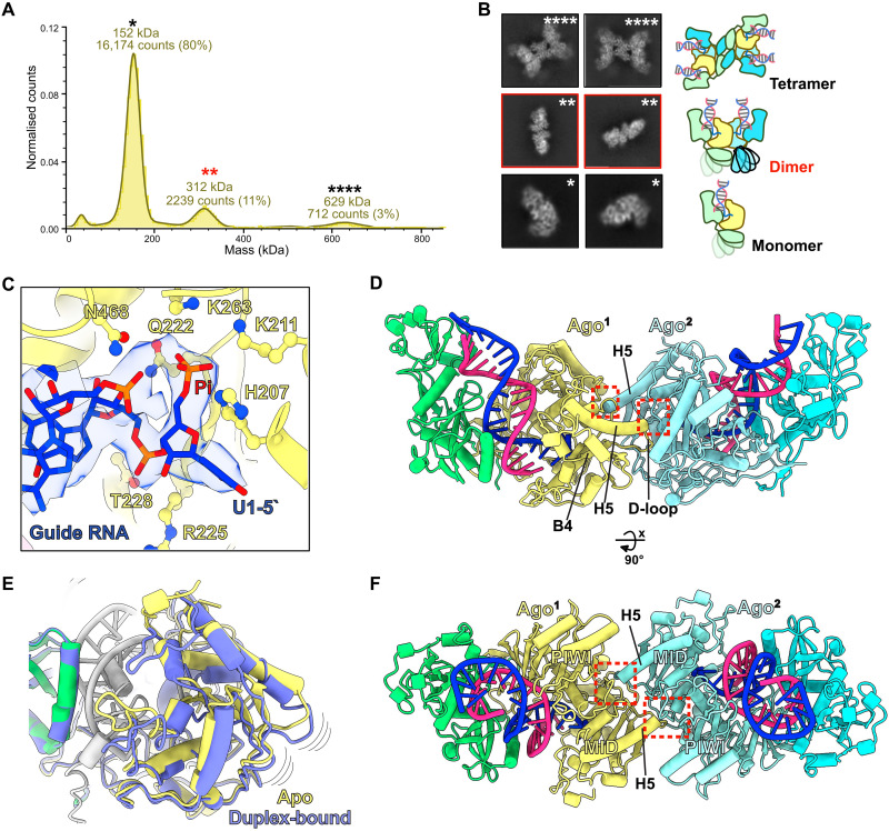
Fig. 2. Assembly of SPARTA dimers upon heteroduplex recognition.
(A) Mass photometry data of the M. polysiphoniae SPARTA (MapSPARTA) incubated with guide RNA (gRNA) and target DNA (tDNA) heteroduplex. The asterisks show molecular weights corresponding to a monomer (*), dimer (**), and tetramer (****). The red asterisks highlight the dimer. (B) Representative two-dimensional class averages of monomer, dimer, and tetramer MapSPARTA. (C) Cartoon and density representation of the 5′ phosphorylated gRNA interacting with residues of the middle (MID) pocket of prokaryotic argonaute (pAgo). (D and F) The structure of dimer state-1 with red dashed boxes indicates the dimerization interface where the C terminus of helix 5 interacts with a pocket formed between the Argonaute (Ago) MID and P-element Induced WImpy (PIWI) domains. (E) Overlay of the Ago MID domain structures in the apo and heteroduplex-bound complexes shows Ago’s structural change upon duplex recognition.

