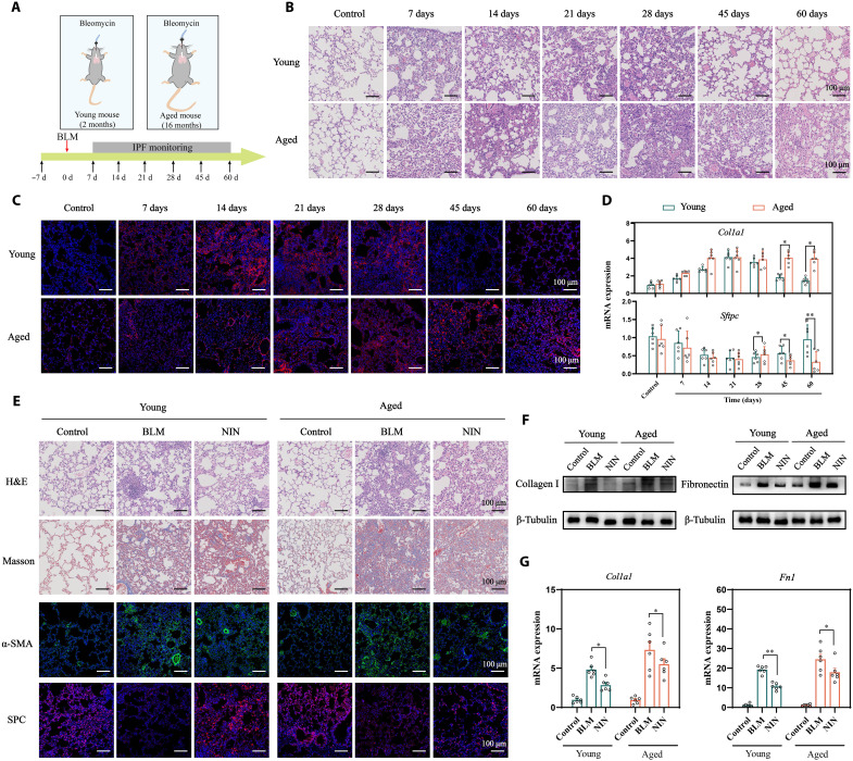
Fig. 2. PF progression in young and aged mice.
(A) Schematic illustration of the study design. H&E staining (B) and IF staining of collagen I (C) in the lungs of young and aged mice challenged with BLM (n = 6). (D) RNA expressions of Col1a1 and Sftpc in the lung of BLM-treated young and aged mice. (E) H&E, Masson, and IF staining of the lungs of young and aged mice treated with NIN (n = 6). WB (F) and qPCR (G) analysis of collagen I and fibronectin (n = 6). Data are means ± SEM. *P < 0.05 and **P < 0.01.

