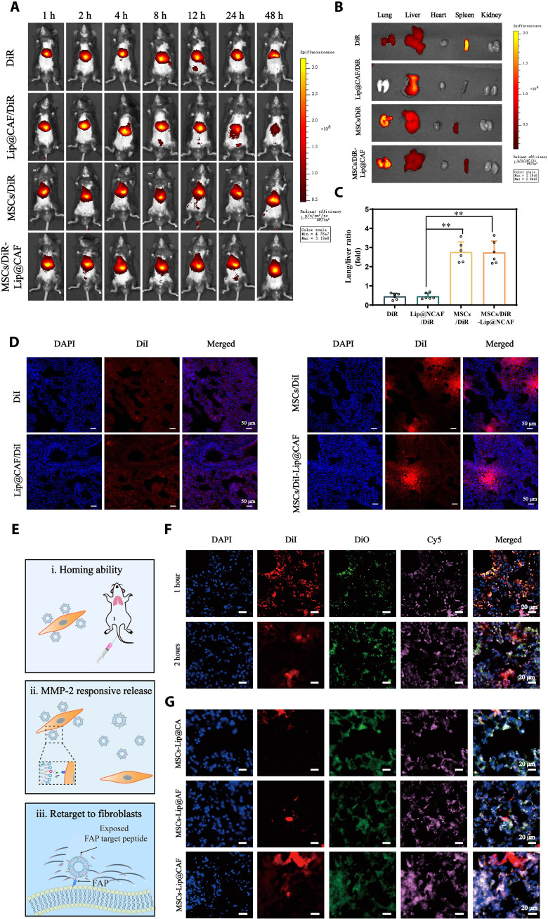
Fig. 5. Homing ability and behavior of MSCs-Lip@NCAF in vivo.
(A) In vivo images of the mice that were intravenously injected with free DiR, DiR-labeled Lip@CAF, MSCs, and MSCs-Lip@CAF (n = 6). (B) Ex vivo images of major organs collected after 12 hours after injection (n = 6). (C) Quantification of the fluorescence intensity of lung/liver (n = 6). (D) Fluorescence imaging of lung sections after the injection of free DiI, DiI-labeled Lip@CAF, MSCs, and MSCs-Lip@CAF for 12 hours. (E) Illustration of the behavior of MSCs-Lip@NCAF in vivo. (F) Fluorescence imaging of the lungs of mice treated with double-labeled MSCs-Lip@CAF (MSCs labeled with DiI and Lip@CAF labeled with DiO) for 1 and 2 hours. (G) IF analysis of lung sections after mice were injected with MSCs-Lip@CA, MSCs-Lip@AF, and MSCs-Lip@CAF (DiO-labeled Lip@CA, Lip@AF, and Lip@CAF, as well as DiI-labeled MSCs) for 4 hours. Data are means ± SD. **P < 0.01.

