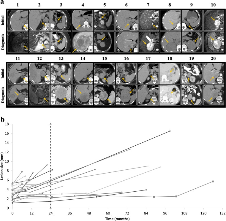
Fig. 1.
a All tumors (yellow arrows), initial imaging (upper panels) and final imaging (lower panels) at time of diagnosis. b Growth of tumors from initial tumor to final size (considering intermediates where available). Dashed line shows 24 month mark, which is the recommended follow-up by several guidelines

