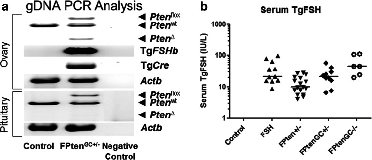
Fig. 1.
a Whole ovary genotyping showed the presence of floxed Pten and the Tg.AMH. Cre-loxP-mediated Pten deletion. In contrast, pituitary had no detectable Pten deletion. Genotyping wild type animals produced the expected PCR products for normal Pten introns. Transgenic Cre and Tg.FSH (β-subunit) screening produced the expected PCR products, and the beta-actin PCR product confirmed the presence of genomic DNA in all samples. b Serum human (Tg)FSH levels were measured using a species-specific immunoassay. TgFSH levels were significantly increased in all Tg females when compared to non-Tg controls (two-way ANOVA G, p < 0.001; FSH, p < 0.001; GxFSH, p < 0.001). Data presented as raw data plotted with median

